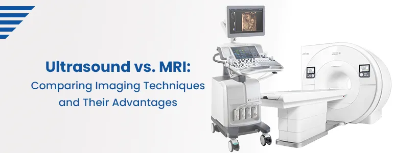
Ultrasound vs. MRI: Comparing Imaging Techniques and Their Advantages

Medical imaging plays a crucial role in diagnosing and monitoring various health conditions. Among the many imaging techniques available today, ultrasound (US) and magnetic resonance imaging (MRI) are widely used modalities. Both methods serve distinct purposes and have their unique advantages. This blog aims to compare these imaging techniques and shed light on their benefits in clinical practice.
Ultrasound Imaging:
Ultrasound imaging, or sonography, utilizes sound waves to create real-time images of the body's internal structures. It is a non-invasive and radiation-free technique, making it safe for patients of all ages, including pregnant women. Ultrasound devices consist of a transducer that emits high-frequency sound waves into the body, which bounce off tissues and organs, generating echoes that are converted into images.
Advantages of Ultrasound:
Real-time Imaging: One of the most significant advantages of ultrasound is its ability to provide real-time images, allowing healthcare professionals to observe dynamic processes such as blood flow and organ movement.
Portability: Ultrasound machines are generally compact and portable, making them readily available for point-of-care use and in remote locations.
Cost-Effective: Compared to other imaging modalities, ultrasound is relatively affordable, contributing to its widespread accessibility in healthcare facilities.
No Ionizing Radiation: Since ultrasound does not involve ionizing radiation, it can be safely used for repeated examinations without significant health risks.
Safe for Specific Patient Groups: Ultrasound is useful for imaging infants, pregnant women, and individuals with certain health conditions that may contraindicate other imaging methods.
Magnetic Resonance Imaging (MRI):
MRI is a powerful imaging technique that uses a strong magnetic field and radio waves to create detailed cross-sectional images of the body's internal structures. It provides excellent soft tissue contrast and is particularly valuable for imaging the brain, spinal cord, joints, and organs.
Advantages of MRI:
Superior Soft Tissue Visualization: MRI reveals intricate details of soft tissues, such as the brain, muscles, tendons, and ligaments, making it invaluable for detecting abnormalities and diseases.
Multi-Planar Imaging: MRI can produce images in various planes (sagittal, coronal, and axial), enabling a comprehensive assessment of anatomical structures from different perspectives.
Non-Invasive and Versatile: MRI is a non-invasive procedure that does not involve ionizing radiation, making it safe for patients of all ages and for repeated examinations when necessary.
Functional and Metabolic Imaging: Advanced MRI techniques like diffusion-weighted imaging (DWI) and magnetic resonance spectroscopy (MRS) allow the assessment of tissue function and metabolism.
Improved Contrast Agents: The development of specialized contrast agents has further enhanced MRI's capabilities, enabling the visualization of specific tissues and abnormalities.
Conclusion:
In conclusion, ultrasound and MRI are valuable imaging techniques with distinct advantages. Ultrasound's real-time imaging, portability, and safety make it ideal for various clinical scenarios, especially in emergencies and when monitoring specific patient groups. On the other hand, MRI's superior soft tissue visualization, multi-planar imaging, and functional assessment capabilities make it indispensable for diagnosing complex conditions and studying neurological disorders.
Ultimately, the choice between ultrasound and MRI depends on the clinical indication, the specific anatomical area being examined, and the patient's unique circumstances. Healthcare professionals should weigh the benefits of each modality to ensure accurate and efficient diagnosis and treatment planning for their patients.
Frequently Asked Questions
Is ultrasound safe during pregnancy?
Yes, ultrasound is considered safe during pregnancy. It uses sound waves instead of ionizing radiation, making it a preferred imaging method for monitoring fetal development. It helps assess the baby's growth, detect abnormalities, and evaluate the placenta and amniotic fluid levels.
Can ultrasound be used for all parts of the body?
Yes, ultrasound can be used to image various parts of the body. It is commonly used for abdominal organs (liver, kidneys, gallbladder), pelvic organs (uterus, ovaries), thyroid gland, heart, blood vessels, and musculoskeletal structures (joints, tendons, muscles).
Are there any limitations to ultrasound imaging?
While ultrasound is a valuable imaging tool, it has some limitations. It may not provide optimal visualization of structures deep within the body, obstructed by air or bone. Additionally, obesity or excess gas in the intestines can hinder image quality. In such cases, MRI may be considered for better assessment.
Can MRI be used in patients with metal implants?
In most cases, MRI can be safely performed on patients with metal implants, but it depends on the type of implant and its compatibility with the magnetic field. Some implants may cause distortions in the MRI image or pose risks to the patient, so informing the healthcare provider about any implants before the procedure is essential.
Which imaging modality is better for evaluating brain conditions?
MRI is generally the preferred imaging modality for assessing brain conditions due to its exceptional soft tissue contrast and ability to provide detailed images of brain structures. It is commonly used to diagnose strokes, tumours, multiple sclerosis, and neurological disorders.
Can ultrasound replace MRI completely?
While ultrasound is a valuable and widely used imaging technique, it cannot wholly replace MRI. MRI's superior soft tissue contrast and ability to visualize deeper structures make it essential for specific diagnoses, such as brain and spinal cord conditions, complex joint pathologies, and evaluating soft tissue tumours.
Is MRI suitable for patients with claustrophobia?
Patients with claustrophobia may find the confined space inside an MRI scanner uncomfortable. However, many modern MRI centres offer open MRI machines or sedation options for patients who experience anxiety or claustrophobia during the procedure.
How long does an ultrasound or MRI examination typically take?
An ultrasound or MRI examination duration varies depending on the body part being imaged and the case's complexity. Generally, ultrasound exams are quicker, taking around 15-30 minutes, while MRI exams can range from 30 minutes to over an hour.
Are there any risks associated with MRI or ultrasound?
Both MRI and ultrasound are considered safe and non-invasive imaging methods. MRI does not use ionizing radiation, making it safe for repeated use. Ultrasound, being radiation-free, is also safe for various patient groups. However, as with any medical procedure, rare allergic reactions to contrast agents used in MRI may occur, and healthcare providers take necessary precautions to minimize risks.
Can both imaging modalities be used together for a more comprehensive evaluation?
Yes, in some cases, ultrasound and MRI may complement each other. For instance, ultrasound can guide needle biopsies or injections accurately, while MRI provides a detailed view of the target area for better diagnosis and treatment planning.
Book Appointment
Our Locations Near You in Hyderabad
3KM from Banjara Hills
1.9KM from Yusufguda
3KM from Madhura Nagar
5KM from Shaikpet