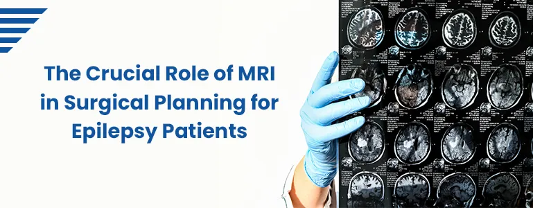
The Crucial Role of MRI in Surgical Planning for Epilepsy Patients

Living with epilepsy can be a challenging journey, but advancements in medical technology have provided new avenues for effective treatment. One such advancement is MRI Epilepsy Protocol for epilepsy patients. In this blog, we'll explore how MRI plays a pivotal role in guiding surgical interventions, enhancing the precision of procedures, and ultimately improving the quality of life for individuals with epilepsy.
Home Sample Collection
Understanding Surgical Options for Epilepsy
When epilepsy cannot be adequately controlled through medications, surgical intervention becomes a viable option. Surgical procedures for epilepsy aim to identify and remove the specific brain regions responsible for seizures. However, pinpointing these regions accurately is a complex process that requires detailed insights into the brain's structure and function. This is where MRI steps in as an invaluable tool.
Role of MRI in Surgical Planning
High-Resolution Structural Imaging: Advanced MRI techniques provide detailed structural images of the brain. These images offer a roadmap for surgeons, allowing them to identify the precise location of abnormal brain tissue, lesions, or structural anomalies that might be causing seizures.
Functional MRI (fMRI): Functional MRI is used to map brain activity by measuring changes in blood flow. This information is crucial for identifying regions of the brain that are actively involved in critical functions like speech, movement, and sensory processing. Surgeons can use this data to avoid damaging these functional areas during surgery.
Diffusion Tensor Imaging (DTI): DTI is used to visualize the pathways of nerve fibers in the brain. In epilepsy surgery, it helps surgeons understand the connectivity between different brain regions, enabling them to plan surgeries that minimize disruptions to essential neural networks.
3D Imaging and Visualization: Modern MRI technology allows for the creation of three-dimensional images of the brain. Surgeons can use these images to simulate the surgery, identifying potential challenges and optimizing their approach before the actual procedure.
The Surgical Process Enhanced by MRI
Preoperative Assessment: MRI provides detailed information about the location and extent of the epileptic focus, guiding neurosurgeons in determining the feasibility of surgery and the potential risks involved.
Defining Surgical Targets: By overlaying structural, functional, and connectivity information from various MRI techniques, surgeons can precisely define the areas to be removed or treated, minimizing damage to healthy brain tissue.
Intraoperative Navigation: Some advanced surgical suites are equipped with intraoperative MRI scanners. These allow surgeons to confirm the accuracy of their surgical plan in real-time, making adjustments if necessary during the procedure.
Postoperative Evaluation: Follow-up MRI scans are conducted after surgery to assess the success of the procedure, monitor healing, and identify any complications that may arise.
Benefits and Future Directions
The integration of MRI into epilepsy surgical planning has transformed the landscape of epilepsy treatment. Patients who once lived with debilitating seizures can now experience significant improvements in their quality of life. As technology continues to evolve, we can expect even more refined imaging techniques, enhanced data visualization, and increased precision in surgical interventions.
Conclusion
The role of MRI in surgical planning for epilepsy patients cannot be overstated. This advanced imaging technology empowers neurosurgeons to make informed decisions, reduce surgical risks, and optimize outcomes for individuals living with epilepsy. As we move forward, the synergy between medical expertise and cutting-edge MRI techniques holds the promise of brighter days for epilepsy patients and their families.
Frequently Asked Questions
What is the role of MRI in surgical planning for epilepsy patients?
MRI (Magnetic Resonance Imaging) plays a fundamental role in epilepsy surgery planning by providing detailed images of the brain's structure, functional activity, and connectivity. These images aid neurosurgeons in precisely identifying seizure-causing regions, preserving essential brain functions, and mapping out surgical strategies.
How does MRI help identify the regions causing seizures?
Advanced MRI techniques, including high-resolution structural imaging and functional MRI (fMRI), help locate abnormal brain tissue, lesions, or anomalies responsible for seizures. By visualizing these areas, neurosurgeons can create a targeted surgical plan to remove or treat the epileptic focus.
What is functional MRI (fMRI), and how does it contribute to surgical planning?
Functional MRI measures changes in blood flow related to brain activity. This information is crucial for mapping areas responsible for vital functions like movement and speech. Surgeons use fMRI data to avoid damaging these areas while addressing the seizure focus.
How does diffusion tensor imaging (DTI) contribute to surgical planning?
Diffusion tensor imaging (DTI) helps visualize nerve fiber pathways in the brain. For epilepsy surgery, DTI provides insights into the connectivity between brain regions. Surgeons can use this information to minimize disruptions to essential neural networks during surgery.
Can MRI help in preserving important brain functions during surgery?
Yes, MRI helps neurosurgeons identify and preserve crucial brain functions. By combining structural, functional, and connectivity data, surgeons can accurately define areas to be treated while minimizing damage to functional regions, like those responsible for speech and movement.
How is MRI used during the actual surgery?
In some advanced surgical suites, intraoperative MRI scanners are available. These scanners allow surgeons to verify the accuracy of their surgical plan in real-time. They can also make adjustments during the procedure to ensure optimal outcomes and reduce the risk of complications.
What happens after surgery? Does MRI play a role in follow-up care?
After surgery, follow-up MRI scans are conducted to evaluate the success of the procedure, monitor healing, and detect any potential complications. These postoperative MRI images provide valuable information about the patient's recovery and the effectiveness of the surgery.
Book Your Slot
Our Locations Near You in Hyderabad
3KM from Banjara Hills
1.9KM from Yusufguda
3KM from Madhura Nagar
5KM from Shaikpet
Profiles
- Cardiac Risk Profile
- Pituitary marker Profile
- Rheumatoid Arthritis Profile
- Dengue Fever Panel
- Lung Cancer Panel 1 Complete Molecular
- Gastroenteritis Screening Panel
- Thyroid Profile (T3,T4,TSH), Serum
- Pancreatic Marker Profile
- STD profile
- Androgen Profile
- Lipid Profile, Serum
- Pancreatic(acute)Profile
- PCOD Profile
Radiology
Pathology Tests
- Glucose Fasting (FBS),Sodium Fluoride Plasma
- Creatinine, Serum
- Glycosylated Hemoglobin (HbA1C)
- Vitamin B12 (Cyanocobalamin), Serum
- Thyroid Stimulating Hormone (TSH) Ultrasensitive, Serum
- Complete Urine Examination (CUE), Urine
- Liver Function Test (LFT),Serum
- Dengue (IgG & IgM), Serum
- Dengue Antigen (Ns1) Rapid, Serum
- C-Reactive Protein (CRP), Serum
- Widal (Slide Method), Serum
- Total IgE, Serum


