Your Cart
Your cart is empty

Looks like you haven't added any test / checkup to your cart

Add Test / Checkup

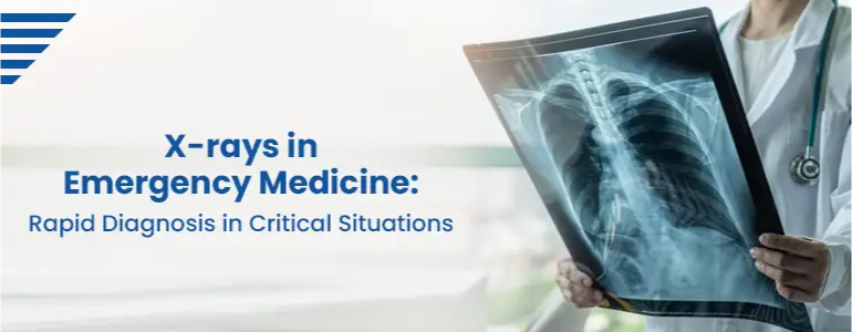
X-rays in Emergency Medicine: Rapid Diagnosis in Critical Situations

X-rays in Emergency Medicine: Rapid Diagnosis in Critical Situations

In emergency medicine, the ability to make fast and accurate diagnoses can mean the difference between life and death. During critical situations, medical professionals rely on various diagnostic tools, and X-rays have become a cornerstone in emergency medical imaging. X-rays provide immediate visual insights into the internal structures of the human body, allowing emergency physicians to swiftly assess injuries and conditions that require urgent intervention. In this blog, we will explore the vital role of X-rays in emergency medicine, their advantages, and how they enable rapid diagnosis in critical situations.

The Importance of X-rays in Emergency Medicine

X-ray imaging has been an indispensable tool in medicine for over a century. Its use in emergency medicine is particularly crucial due to the need for quick and accurate assessments in time-sensitive situations. X-rays can help diagnose a wide range of conditions, from bone fractures and dislocations to life-threatening conditions such as pneumothorax, pulmonary edema, and abdominal trauma. The ability to identify these conditions rapidly allows emergency physicians to prioritize and initiate appropriate treatments promptly.

Advantages of X-rays in Emergency Medicine

  • Speed and Efficiency:X-rays are non-invasive and produce immediate results, making them ideal for fast-paced emergency situations. Physicians can obtain X-ray images within minutes, allowing for real-time assessments and swift decision-making.
  • Critical Information:X-ray images provide essential information about fractures, dislocations, foreign objects, and soft tissue abnormalities that may not be visible through physical examination alone. This critical information guides treatment plans and helps avoid potential complications.
  • Accessibility:X-ray machines are readily available in most emergency departments, ensuring rapid access to this diagnostic tool. The convenience of on-site X-ray imaging contributes to efficient patient management and reduces the need for patient transfers to other departments or facilities.
  • Guiding Procedures:In emergency medicine, X-rays often serve as a guide for procedures such as joint reductions, chest tube placements, and central line insertions. Real-time imaging helps ensure the accurate and safe execution of these interventions.
  • Minimizing Radiation Exposure:X-ray technology has advanced significantly, allowing for the use of lower radiation doses without compromising image quality. This minimizes patient radiation exposure, particularly important in situations where multiple imaging studies may be required.

X-rays in Specific Emergency Situations

  • Trauma and Fractures:In cases of trauma, such as motor vehicle accidents or falls, X-rays are instrumental in diagnosing fractures, dislocations, and soft tissue injuries. Immediate identification of these injuries enables timely splinting, casting, or surgical intervention, reducing the risk of further damage.
  • Chest Injuries:X-rays of the chest are essential in evaluating patients with suspected rib fractures, pneumothorax, hemothorax, or pulmonary contusions. Rapid identification of these conditions guides appropriate management, such as chest tube insertion for pneumothorax.
  • Abdominal Trauma:X-rays play a role in diagnosing abdominal injuries, identifying free air in the abdomen (pneumoperitoneum) suggestive of organ perforation. They can also reveal foreign objects or potential sources of internal bleeding.
  • Respiratory Emergencies:X-rays help diagnose conditions such as pneumonia, pleural effusions, and pulmonary edema, which may present with acute respiratory distress. Rapid diagnosis allows timely initiation of appropriate treatments.
  • Joint Dislocations:X-rays are crucial in diagnosing joint dislocations, providing guidance for reduction procedures to realign the joint and relieve pain.
  • Foreign Body Ingestion or Aspiration:X-rays can identify swallowed or aspirated foreign bodies, aiding in determining the appropriate management, such as removal or further evaluation.

Limitations and Considerations

While X-rays are invaluable in emergency medicine, it is essential to recognize their limitations and consider potential risks:

  • Radiation Exposure:Despite advances in technology, X-ray imaging still involves some level of ionizing radiation. Emergency physicians must carefully weigh the risks and benefits of each X-ray study, especially in vulnerable populations like pregnant women and children.
  • Limited Soft Tissue Detail:X-rays primarily excel in imaging bones, but they have limited capabilities in providing detailed views of soft tissues like muscles and organs. Complementary imaging modalities like ultrasound or CT scans may be required for a comprehensive evaluation.
  • Overutilization:In the fast-paced emergency setting, there is a risk of overutilization of X-rays due to the desire for quick answers. Clinicians should adhere to evidence-based guidelines to ensure appropriate and judicious use of imaging studies.

Conclusion

X-rays have become an invaluable tool in emergency medicine, providing rapid and critical information in time-sensitive situations. Their ability to diagnose fractures, dislocations, pneumothorax, and various other conditions allows emergency physicians to make informed decisions and initiate prompt treatments. While X-rays have revolutionized emergency medicine, healthcare providers must exercise caution in their usage to minimize radiation exposure and ensure appropriate diagnostic imaging. As technology continues to evolve, we can expect even more advanced X-ray systems that further enhance patient care and outcomes in emergency medicine.

Home Sample Collection Process

1
Book your convenient slot
2
Sample Collection by Phlebotomist
3
Reporting of the sample at lab
4
Download Reports
Download Reports

Note: Home Sample Collection is only for Pathology lab tests.

Frequently Asked Questions

X-rays play a critical role in emergency medicine as they provide immediate visual insights into the internal structures of the body. In critical situations, when swift and accurate diagnoses are crucial, X-rays enable emergency physicians to quickly assess injuries, fractures, dislocations, and life-threatening conditions. Their immediate availability and non-invasive nature make X-rays indispensable in time-sensitive scenarios, guiding prompt treatment decisions.

X-rays are particularly effective in diagnosing fractures and bone injuries during emergencies. They allow physicians to visualize bone structures, identify fractures, and determine the extent and alignment of the injury. This information is vital for determining appropriate treatments, such as splinting, casting, or surgery, to prevent further complications.

X-rays are routinely used in various emergency situations, including traumatic injuries from accidents or falls, suspected rib fractures, joint dislocations, chest injuries, abdominal trauma, foreign body ingestion or aspiration, and acute respiratory distress. In all these scenarios, X-rays provide immediate insights, guiding emergency physicians in their decision-making process.

While X-rays are valuable in emergencies, the potential risk of radiation exposure should be considered. Advanced X-ray technology has significantly reduced radiation doses, making them safer. However, it is essential for healthcare providers to adhere to appropriate protocols and consider the benefits versus the risks when ordering X-ray studies, especially in vulnerable populations like pregnant women and children.

X-rays serve as invaluable guides for various emergency procedures. For instance, they aid in the reduction of joint dislocations, accurate placement of chest tubes for pneumothorax, and the insertion of central lines. Real-time imaging during these procedures ensures their safe and precise execution, minimizing potential complications.

Yes, X-rays can identify various conditions beyond fractures in emergency medicine. They can help diagnose pneumothorax (collapsed lung), hemothorax (blood in the chest cavity), pneumonia, pleural effusions, abdominal injuries, foreign body ingestion, and aspiration. X-rays provide essential information to evaluate patients with respiratory distress or acute abdominal pain, guiding appropriate and timely treatments.

To ensure judicious use of X-rays in emergency medicine, healthcare providers should follow evidence-based guidelines for diagnostic imaging. Clinicians should carefully weigh the benefits and risks of each X-ray study, considering alternative imaging modalities when appropriate. Additionally, effective communication among the healthcare team can aid in determining the most appropriate diagnostic approach for each patient, avoiding unnecessary radiation exposure when possible.

Book Your Slot
Locations Near You in Hyderabad
  • 4KM from Madhapur
  • 3KM from Banjara Hills
  • 1.9KM from Yusufguda
  • 3KM from Madhura Nagar
  • 5KM from Shaikpet