Your Cart
Your cart is empty

Looks like you haven't added any test / checkup to your cart

Add Test / Checkup

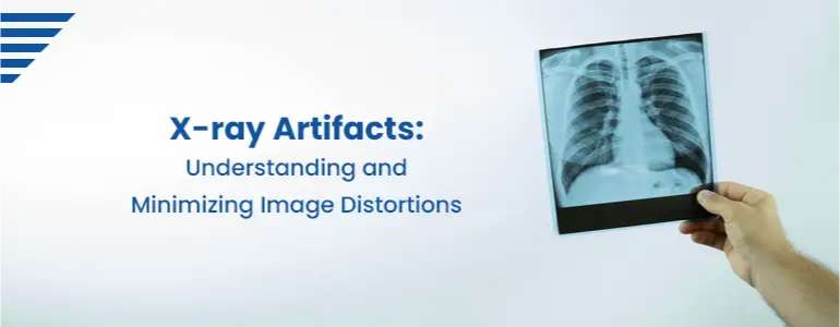
X-ray Artifacts: Understanding and Minimizing Image Distortions

X-ray Artifacts: Understanding and Minimizing Image Distortions

X-ray artifacts are unwanted, unexpected, and often misleading features or structures that appear on X-ray images. They are not representations of the anatomy being imaged and can result from various factors during the imaging process. These artifacts can hinder accurate interpretation and diagnosis, leading to potential errors in patient care.

Home Sample Collection Process

1
Book your convenient slot
2
Sample Collection by Phlebotomist
3
Reporting of the sample at lab
4
Download Reports
Download Reports

Note: Home Sample Collection is only for Pathology lab tests.

Causes of X-ray artifacts include:

  • Patient-related Factors: Artifact can arise due to patient movement during the exposure, improper positioning, or the presence of metal objects (e.g., jewellery, dental fillings, implants) within the body.
  • Technical Factors: Errors in X-ray machine calibration, incorrect exposure settings, or issues with the X-ray detector can introduce artifacts into the image.
  • Processing Artifacts: Improper handling or processing of X-ray films or digital images can cause artifacts.
  • Scatter Radiation: Scatter radiation occurs when X-rays interact with tissues, diverting their path and creating unwanted effects on the final image.
  • Grid Artifacts: Using grids to reduce scatter can lead to grid lines or MoirĂ© patterns on the image if not aligned correctly.
  • Beam Hardening: This occurs when low-energy X-rays are absorbed by the body, leaving behind higher-energy X-rays that can create streaking artifacts.
  • Metal Artifacts: Metallic objects (e.g., prosthetics, screws, pacemakers) can cause streaks or shadows on the X-ray due to their high density and ability to absorb X-rays.
  • Motion Artifacts: Inadequate patient cooperation or involuntary motion can result in blurring or distortion of the image.
  • Cassette Artifacts: Dust, scratches, or damage to the X-ray cassette can lead to artifacts on the image.

Addressing artifacts is crucial in obtaining high-quality X-ray images for accurate diagnosis and treatment planning. Radiologic technologists and radiologists are trained to identify and mitigate these artifacts, and technological advancements in X-ray imaging equipment have helped reduce their occurrence over time.

Types of X-ray Artifacts:

  • Patient Motion Artifacts: Explaining how patient movement during exposure can lead to blurring or ghosting in the image.
  • Beam Hardening Artifacts: Discussing the impact of varying tissue densities on X-ray beam attenuation and the resulting artifacts.
  • Scatter Radiation Artifacts: Understanding the effects of scattered X-rays on image quality and how it can be managed.
  • Metal Artifacts: Explaining how metallic objects within the body cause streaks or shadows on X-rays and their impact on image interpretation.

Impact on Diagnosis:

  • The Importance of Artifact Identification:Emphasizing recognizing artifacts and distinguishing them from proper anatomical structures.

  • Potential Misdiagnoses:Discuss the consequences of misinterpreting artifacts as pathologies, leading to unnecessary treatments or missed diagnoses.

Strategies to Minimize X-ray Artifacts:

  • Optimal Patient Positioning: Highlighting the importance of proper positioning to minimize motion artifacts and enhance image clarity.

  • Beam Hardening Compensation: Exploring methods to address beam hardening, such as using advanced X-ray filters or energy-based corrections.

  • Scatter Radiation Management: Discussing anti-scatter grids and advanced collimation techniques to reduce scatter artifacts.

  • Metal Artifact Reduction Techniques: Introducing methods like metal artifact reduction software and dual-energy X-ray imaging.

Advancements in X-ray Technology:

Digital Radiography (DR) and Computed Radiography (CR): Exploring how digital X-ray technologies have reduced artifacts and improved image quality.

Dual-Energy X-ray Imaging: Highlighting the benefits of dual-energy techniques in mitigating artifacts caused by metal objects.

Conclusion:

X-ray artifacts can significantly impact the diagnostic quality of X-ray images, leading to potential misdiagnoses and compromised patient care. Understanding the different types of antiques and their causes is essential for radiologic technologists and radiologists. By implementing strategies to minimize artifacts and embracing advancements in X-ray technology, medical professionals can ensure the production of high-quality diagnostic images, facilitating accurate diagnoses and improved patient outcomes.

Frequently Asked Questions

X-ray artifacts are unwanted image distortions that appear on X-ray images, hindering accurate diagnosis. They can arise from various factors, such as patient motion, technical errors, or the presence of metal objects. Artifacts can be misleading, leading to potential misdiagnoses and compromised patient care.

Patient motion artifacts occur when the patient moves during the X-ray exposure. This movement can result in blurring or ghosting in the image, making it challenging to accurately assess anatomical structures.

Beam hardening occurs when X-rays pass through different tissue densities, leading to variations in their energy levels. This phenomenon can cause streaking artifacts in the image, particularly around dense structures like bones.

Scatter radiation occurs when X-rays interact with tissues and change direction. Excessive scatter radiation can reduce image contrast and clarity, leading to poor image quality and difficulty identifying fine details.

Yes, metal objects such as prosthetics, screws, or dental fillings can cause streaks or shadows on X-ray images due to their high density. These artifacts can obscure nearby anatomical structures and impact image interpretation.

Imaging technicians play a crucial role in minimizing X-ray artifacts. Proper patient positioning, communication, and cooperation with patients to minimize motion are essential steps to reduce motion artifacts. They can also adjust exposure settings and use collimation techniques to manage scatter radiation.

Advanced X-ray equipment, such as digital radiography (DR) and computed radiography (CR), have improved image processing capabilities. They can help reduce artifacts through image enhancement algorithms and software tools that compensate for beam hardening and scatter radiation.

Dual-energy X-ray imaging involves capturing images at different energy levels, which allows for better tissue differentiation and artifact reduction. It is particularly effective in minimizing artifacts caused by metal objects.

Experienced radiologists can differentiate between true anatomical structures and artifacts. Radiology training includes recognizing and distinguishing various artifacts from actual pathologies, ensuring accurate diagnosis and treatment planning.

Book Your Slot
Locations Near You in Hyderabad
  • 4KM from Madhapur
  • 3KM from Banjara Hills
  • 1.9KM from Yusufguda
  • 3KM from Madhura Nagar
  • 5KM from Shaikpet