Your Cart
Your cart is empty

Looks like you haven't added any test / checkup to your cart

Add Test / Checkup

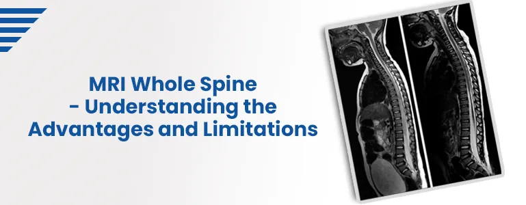
MRI Whole Spine - Understanding the Advantages and Limitations

MRI Whole Spine - Understanding the Advantages and Limitations

Magnetic Resonance Imaging (MRI) has revolutionized medical diagnostics, allowing for non-invasive and detailed visualization of internal structures. One significant application of MRI is the examination of the entire spine, known as "MRI Whole Spine." This advanced imaging technique offers a comprehensive view of the spinal column, aiding in diagnosing and managing various spinal conditions. However, like any medical procedure, an MRI of the Whole Spine has advantages and limitations that are essential to understand.

Advantages of MRI Whole Spine

  • Comprehensive Visualization: MRI Whole Spine provides a holistic view of the spinal cord, from the cervical to the sacral regions. This is crucial for identifying issues that might span multiple spine segments, enabling accurate diagnosis and treatment planning.
  • Non-Invasiveness: Unlike traditional methods like myelography or CT myelography, which involve injecting contrast agents into the spinal canal, MRI Whole Spine is non-invasive. It avoids the need for needles or exposure to ionizing radiation.
  • Detailed Soft Tissue Imaging: MRI is particularly adept at imaging soft tissues like nerves, intervertebral discs, ligaments, and blood vessels. This capability is essential for diagnosing herniated discs, spinal stenosis, tumours, and other conditions that affect these structures.
  • Multi-Planar Imaging: MRI Whole Spine can produce images in multiple planes (sagittal, coronal, and axial), allowing radiologists to visualize the spine from different angles. This aids in obtaining a more accurate assessment of spinal abnormalities.
  • Improved Contrast Resolution: MRI offers excellent contrast resolution, clearly differentiating between various tissues. This is particularly advantageous when distinguishing between normal and pathological tissues.

Limitations of MRI Whole Spine

  • Limited Accessibility for Certain Patients: Some patients, such as those with claustrophobia, obesity, or certain medical implants (e.g., pacemakers), may not be suitable candidates for MRI due to the confined space of the MRI machine or potential safety concerns.
  • Time-Consuming: MRI Whole Spine can be time-consuming, often requiring patients to remain within the machine for an extended period. This can be challenging, especially for patients with difficulty lying still or in pain.
  • Cost: MRI is generally more expensive than other imaging modalities, which might limit its accessibility for some individuals or healthcare systems.
  • Artefact Interference: Metallic implants, foreign bodies, or motion during the scan can lead to artefacts in the images, potentially obscuring the diagnostic quality of the scan.
  • Limited Sensitivity to Bone Abnormalities: While MRI is excellent at imaging soft tissues, it may not be as sensitive as CT scans for detecting specific bone abnormalities, fractures, or degenerative changes in bone.

Conclusion

MRI Whole Spine is a powerful tool for diagnosing and understanding spinal conditions. Its non-invasive nature and ability to provide detailed soft tissue images make it a preferred choice in many cases. However, it's essential to recognize its limitations, such as accessibility challenges, time requirements, and potential artefacts. The decision to use MRI Whole Spine should always be based on a careful assessment of the patient's condition, clinical needs, and available resources, ensuring the best possible outcome for accurate diagnosis and effective treatment planning.

Home Sample Collection Process

1
Book your convenient slot
2
Sample Collection by Phlebotomist
3
Reporting of the sample at lab
4
Download Reports
Download Reports

Note: Home Sample Collection is only for Pathology lab tests.

Frequently Asked Questions

MRI Whole Spine is a comprehensive imaging technique that captures detailed images from the cervical to the sacral sections of the spinal column. Unlike traditional methods that may involve injections of contrast agents or ionizing radiation, MRI uses good soft tissue vision and is non-invasive since it creates images using radio waves and magnetic fields.

MRI Whole Spine offers several benefits, including comprehensive visualization of the entire spinal cord, detailed imaging of soft tissues such as nerves and discs, multi-planar imaging for better assessment, and high contrast resolution for accurate tissue differentiation. It is also non-invasive and avoids the use of radiation.

While MRI of the Whole Spine is a valuable diagnostic tool, it might only suit some. Patients with claustrophobia, obesity, or certain medical implants, like pacemakers, might face limitations. Additionally, the time-consuming nature of the procedure and potential cost considerations can also be factors to consider.

MRI Whole Spine is highly effective in diagnosing various spinal conditions. It provides:

Yes, there are limitations to consider. MRI Whole Spine might not be feasible for patients with certain medical conditions or implants. The procedure can be time-consuming, and its cost might be a limiting factor for some individuals or healthcare systems. Additionally, artefacts from metallic implants or motion during the scan can affect image quality.

While MRI Whole Spine excels in visualizing soft tissues, it may not be as sensitive as CT scans when it comes to detecting specific bone abnormalities, fractures, or degenerative changes in bone. CT scans are better suited for evaluating bone structures.

Patients should follow any preparation instructions provided by their healthcare provider. This might include removing metal objects, wearing comfortable clothing, and informing the medical team about any medical implants or conditions that could affect the procedure.

Absolutely. MRI Whole Spine can be used to monitor treatment progress for spinal disorders. By comparing images taken before and after treatment, medical professionals can assess the effectiveness of interventions and make any necessary adjustments.

In some cases, sedation might be considered for patients with problems lying still during the procedure. This decision would depend on the patient's age, medical condition, and the medical team's situation assessment.

Healthcare providers will consider the patient's medical history, symptoms, and clinical presentation to determine the most appropriate imaging option. An MRI of the Whole Spine might be recommended if comprehensive visualization of the spinal column and detailed soft tissue images are required.

Book Your Slot
Locations Near You in Hyderabad
  • 4KM from Madhapur
  • 3KM from Banjara Hills
  • 1.9KM from Yusufguda
  • 3KM from Madhura Nagar
  • 5KM from Shaikpet